ONLINE
PHONE
(800) 621-8335
FAX
(312) 464-5600
16
SPRING2017
AMERICANMEDICALASSOCIATION
An invaluable resource forCPT® surgical codingwith in-context
anatomical knowledgeand illustrations! Thispublication from the
AMAcombinesCPTsurgerycodesanddescriptionswithclinicallyand
anatomically significantdrawings from renownedmedical illustrator
FrankH.Netter,MD.
Netter’sAtlasofSurgicalAnatomy forCPT®Coding
isan ideal supplement to theCPTcodebook.Gainanadditional
boostof anatomical knowledge in thecontextofCPTcodesasyou
abstract codes fromphysiciannotesandoperative reports.
Softbound, 8½" x11", 516pages
Item#:OP495015
ISBN: 978-1-62202-030-0
Price: $119.95
AMAMEMBERPRICE:$89.95
eBook
Item#: EB495015
ISBN: 978-1-62202-031-7
Price: $119.95
AMAMEMBERPRICE:$89.95
CodingAtlas, aunique feature thatpresents
anatomydetails related toprocedureswithin
specificCPTcode ranges.
Neurostimulators (Intracranial)
CodingAtlas
An
intracranial
neurostimulator is an electrode array
implanted at apredetermined site to suppress tremors,
seizures, or other
functionaldisorders
.
Bilateral
symptoms and findingsmay require treatmentwith
bilateral electrode arrays.One electrode arraymay be
connected to onepulse generator or receiver, or two
electrode arrays (one on each side of the brain)maybe
required and be connected to one generator or receiver.
The
neurostimulator
insertion/removal codesdonot
include evaluation, testing,programming, or
reprogramming.These services are reportedwith codes
from theMedicine section of theCPT code set.
61850
Twist drill or
burrhole
(s) for implantation of
neurostimulator
electrodes, cortical
61860
Craniectomy
or
craniotomy
for implantation of
neurostimulatorelectrodes, cerebral, cortical
61863
Twist drill,burr hole, craniotomy,or craniectomywith
stereotactic implantation of neurostimulatorelectrode
array in subcortical site (eg, thalamus,globus pallidus,
subthalamic nucleus, periventricular, periaqueductal
gray),without useof intraoperativemicroelectrode
recording; first array
✚
61864
each additionalarray (List separately inaddition to
primary procedure)
61867
Twist drill,burr hole, craniotomy, or craniectomywith
stereotactic
implantationofneurostimulator electrode
array in subcortical site (eg, thalamus, globuspallidus,
subthalamic nucleus, periventricular, periaqueductal
gray),withuse of
intraoperativemicroelectrode
recording
; first array
✚
61868
each additionalarray (List separately inaddition to
primary procedure)
61870
Craniectomy for implantation ofneurostimulator
electrodes, cerebellar, cortical
61880
Revision or removal of intracranial neurostimulator
electrodes
61885
Insertion or replacement of cranial neurostimulator pulse
generator or receiver, direct or inductive coupling;with
connection toa singleelectrode array
61886
with connection to 2 ormore electrode arrays
61888
Revision or removal of cranial neurostimulatorpulse
generator or receiver
FIGURE 9-14.
FunctionalAreas forNeurostimulation
Intracranial
neurostimulation
describes surgicalplacement of an electrical impulseprobe for
therapeutic reasons.For example, a
deepbrain stimulator
(DBS)placed in the globuspallidus
interni (GPi) or subthalamicnucleus (STN)may reduce symptoms ofParkinsondisease, essential
tremor, andmultiple sclerosis. Stimulation of the vagalnerve coming from themedulla oblongata
maybeprescribed forpatientswith epilepsywho are intolerant of antiepilepticdrug therapy or
whose seizure activity cannotbe adequately controlled.
Limbic cingulate
cortex
Thalamus
Pituitary gland
Pons
Supplemental
motor cortex
Medulla oblongata
Frontal
Limbic
Primarymotor cortex
Parietal
Occipital
Precentral sulcus
Paracentral lobule
Somatosensory association cortex
Corpus callosum
Visual association cortex
Calcarine
fissure
Primary visual cortex
Cerebellum
61850—61888
NervousSystem
Netter’sAtlas ofSurgicalAnatomy forCPTCoding
390
~
Moderatesedation
✚
Add-oncode
Modifier51exempt
#
Resequencedcode
NettersAtlas_PASS 5.indb 390
1/7/15 2:18PM
Figurecaptionsassist incode selectionby
providing full descriptionsabout the illustrated
anatomiesandprocedures.
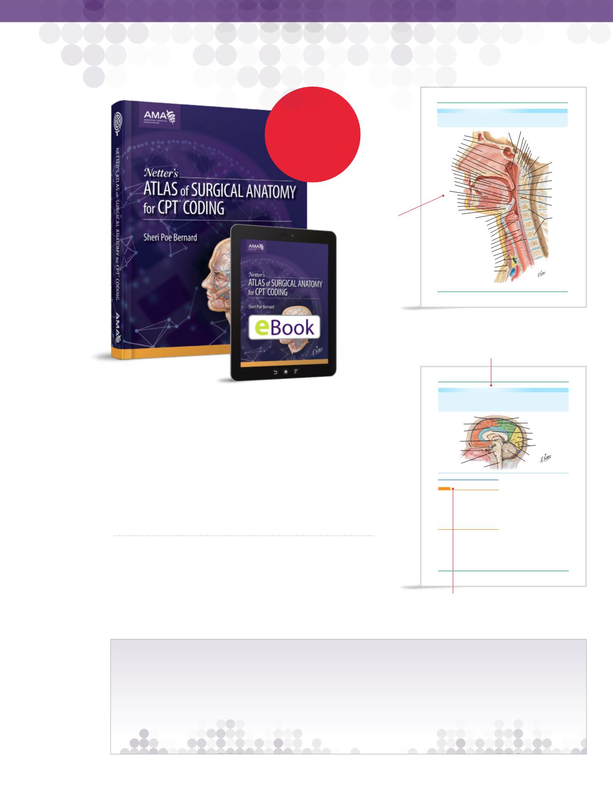
Sella turcica
Frontal sinus
Sphenoidal sinus
Nasal septum
Nasopharynx
Soft palate
Palatine glands
Hard palate
Oral cavity
Incisive canal
Palatine tonsil
Body of tongue
Oropharynx
Foramen cecum
Lingual tonsil
Genioglossus
muscle
Root of tongue
Epiglottis
Mandible
Geniohyoidmuscle
Hyoid bone
Hyoepiglottic ligament
Thyrohyoidmembrane
Laryngopharynx
Laryngeal inlet (aditus)
Thyroid cartilage
Vocal fold
Transverse arytenoidmuscle
Cricoid cartilage
Trachea
Esophagus
Esophagealmuscles
Thyroid gland
Superficial (investing) layer of deep cervical fascia
Pretracheal fascia
Suprasternal space (ofBurns)
Manubrium of sternum
Pharyngeal opening of auditory
(pharyngotympanic, eustachian (tube)
Sphenooccipital synchondrosis
Pharyngeal tonsil
Pharyngeal tubercle of occipital bone
Pharyngeal raphe
Anterior longitudinal ligament
Anterior atlantooccipitalmembrane
Apical ligament of dens
Pharyngeal
constrictor
muscles
Bucco-
pharyngeal
fascia
Retro-
pharyngeal
space
Prevertebral
fascia and
anterior
longitudinal
ligament
Vertebral
bodies
Anterior
arch of
atlas (C1
vertebra)
Densof
axis (C2
vertebra)
C1
C1
C2
C3
C4
C5
C6
C7
T1
FIGURE3-7.
TheTracheaandPharynx
Thepharynx communicateswith the esophagus,nasal cavity,middle ear, and larynx. It isdivided into
three anatomical sites:nasopharynx, oropharynx, and laryngopharynx.The trachea, orwindpipe, is
composed of about20 rings of tough
cartilage
and is lined in
mucosa
.At its base, itdivides into the
right and leftbronchus.A
posterior
view of thepharynx can be seen inFigure 5-6.
136
~
Moderatesedation
✚
Add-oncode
Modifier51exempt
#
Resequencedcode
RespiratorySystem
Netter’sAtlas ofSurgicalAnatomy forCPTCoding
NettersAtlas_PASS 5.indb 136
1/7/15 2:15PM
More than700
individualNetter
illustrations
pairedwith
specificcode
ranges toguide
code selection.
VIDEO
TIONAT
RE.COM
PurchasewithaCPT®2018Professional title to
BUILDYOUROWN
PACKAGE
SAVEABUN
DLE
with


